
Considering a Root Canal or Concerned About One You Already Have?
Many of the patients I meet have already been told by another dentist that a root canal is needed, or have already had a root canal that they are concerned could be infected. Rather than looking for someone to contradict that recommendation, they are rightfully concerned about the possible long-term issues of a root canal. A second-opinion focused exam follows four steps:
- Bring your existing X-rays, reports, and anything you were told about the tooth.
- We examine the tooth and talk with you about your symptoms.
- We determine whether a CBCT scan is needed for a more complete view.
- We review the findings with you and discuss your options.
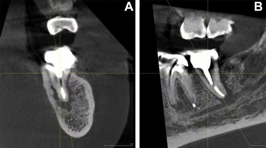
A CBCT (cone beam computed tomography) creates 3D cross-sectional views of the tooth, its roots, and the surrounding bone. All our offices have CBCT scanners on site.
This scan allows us to look closely at the bone surrounding the root, and we recommend root canal treatment only when that bone appears healthy. This is a more cautious approach than many dentists take because regardless of what you are told, there is no way to remove every speck of infectious material from a tooth during a root canal procedure.
Even though a scan shows us how the surrounding bone has responded to inflammation from an infection, it cannot show us bacteria. That is why we never make the decision from an image alone. We consider it alongside your physical exam, tooth testing, and symptoms before recommending a path forward.
Our Philosophy on Root Canals
We are not against every root canal, and we do not believe every root-canal-treated tooth should be removed. But we are more cautious than most.
When a tooth can be restored and the surrounding bone looks healthy, we recommend root canal treatment and refer you to an endodontist who uses a microscope.
For a tooth that has already had a root canal, healthy surrounding bone is reassuring. If imaging shows a persistent or growing lesion, we take that seriously—especially since a tooth with the nerve removed cannot cause pain. In those cases, we are more likely to recommend addressing the root cause of the problem vs. monitoring it year after year.
Why We Take a Cautious Approach
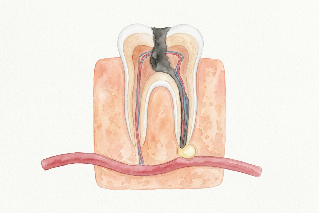
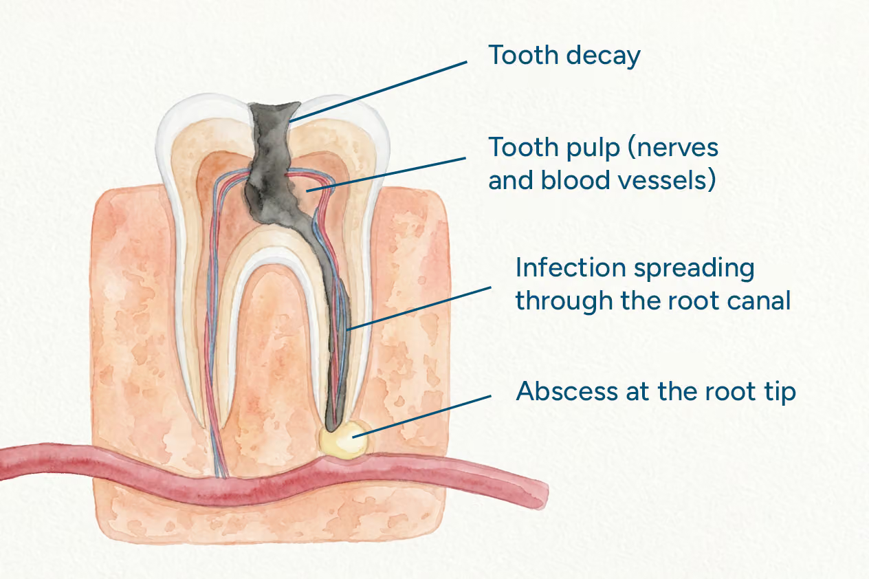
Inside a tooth is less like one straight pipe and more like a maze of microscopic channels. Those channels extend into even smaller spaces called dentinal tubules. Instruments and disinfecting liquids can dramatically reduce bacteria, but they cannot reach the full depth of every tubule. If infected tissue or bacteria remain in those microscopic spaces, they can sustain a low-grade infection long after treatment is complete.
During a root canal, the pulp, including its nerves and blood vessels, is removed from inside the tooth. The tissues around the root remain alive. That surrounding bone becomes our scoreboard: healthy-looking bone suggests healing, while a persistent or growing area of bone change tells us healing is incomplete.
Where We Draw the Line
Apical periodontitis is inflammation in the bone and tissues around the tip of a tooth’s root. In our view, persistent inflammation around a tooth deserves attention even when there is “no pain”.
Retreatment or an apicoectomy can sometimes save the tooth, and an endodontist can evaluate those options. Where our philosophy differs is indefinite watchful waiting. Losing a tooth is a known cost that we can plan for. Carrying an unresolved lesion for ten years is a concern for your long-term health.
If the bone is not healing—or the lesion is growing—we would rather revisit the options than keep adding the same note at every checkup.
The Specialists We Refer To
The endodontists we refer to will not treat a tooth they consider to be a long term health risk to the body. They share our belief that treatment should proceed only when a tooth has a reasonable chance of success, and they perform root canals using specialized equipment such as a microscope, ozone gas, and gentle-wave technology.
“Endodontic infection can have systemic ramifications. It’s imperative to disinfect the canal system as thoroughly as possible. The new technologies such as Gentlewave and Fotona laser are helping to disinfect areas that were not possible to disinfect with traditional root canal methods. Saving your teeth saves your jaw bone, prevents bone microfractures, and sends feedback to your brain because periodontal ligaments around a root canal tooth are still alive. I use ozone water as final irrigant, ozone gas to insufflate the canals and photobiomodulate the area (treat with low-level laser and LED light). Ozone and PBM shift the treatment from disinfection to healing by modulating inflammatory response. It’s also important to follow up and assure healing by having yearly CBCT imaging.”
Dr. Amber Chu, DDS, Puget Sound Endodontics
Where I part ways with some of the profession is watch-and-wait. Some areas around the root tip heal after treatment, and allowing time for that healing is sometimes appropriate. But I am not comfortable watching a persistent or growing lesion year after year simply because the tooth does not hurt.
I would never risk the long term health of one of my own family members, and the same intention is given to our patients at The Airway Dentists.
What a 3D Scan Reveals That Flat X-Rays Miss
A regular dental X-ray is a flat picture of a three-dimensional area. Because roots and bone overlap, dentists take images from more than one angle.
We’ve missed infected teeth on 2D X-rays before, because you cannot rotate the image or fully evaluate every root from a single flat view. When symptoms and X-rays don’t match—or when something still doesn’t add up—a CBCT scan lets us look at the tooth and surrounding bone in cross-sections and better estimate the true size and extent of any lesion.
In my experience, a tooth that looks reassuring on a flat X-ray can look very different in 3D.
What the Numbers Show
A periapical lesion is an area of bone change around the tip of a tooth’s root. It often points to inflammation caused by infection inside the tooth, although an image alone cannot confirm that an active infection is present.
That doesn’t mean every tooth needs a 3D scan. It means that when a flat X-ray leaves a meaningful question unanswered, a CBCT may reveal information that changes the treatment plan.
What Happens After the Scan
If the Surrounding Bone Looks Healthy
If the tooth still needs root canal treatment, we refer you to an endodontist because we do not perform root canals in our offices.
An endodontist is a specialist who treats disease inside the tooth and around its roots. We work with specialists whose philosophy aligns with ours and who provide the level of care we consider appropriate for complex treatment. You are always free to choose your own specialist.
If the Scan Shows a Concerning Area
When a scan shows a significant lesion or cyst-like area around a root—especially in a previously treated tooth—we usually recommend removing the tooth. Our goal is to remove the source of the problem rather than continue treating around it.
That is my philosophy, but it is not the only option. If you decide to keep your tooth, other documented options include:
- Retreatment: Reopening the tooth, removing the old filling material, cleaning the canals again, and resealing them.
- Apicoectomy: A small surgery that removes the affected tissue around the root end, along with the tip of the root.
Both treatments can have favorable results, although outcomes vary. An endodontist can help determine whether either option is appropriate for the tooth.
Already Have a Root Canal? Why Problems Can Be Silent
Root canal treatment removes the inflamed or infected pulp from the tooth, which includes the nerve inside it. The tissue surrounding the root still has nerves, so a failing treated tooth may ache, swell, or hurt when you bite.
Not every problem causes symptoms, though. Some changes remain quiet until imaging reveals them. Pain is an imperfect warning system. A quiet tooth is not necessarily a healthy tooth.
What I See and What the Research Shows
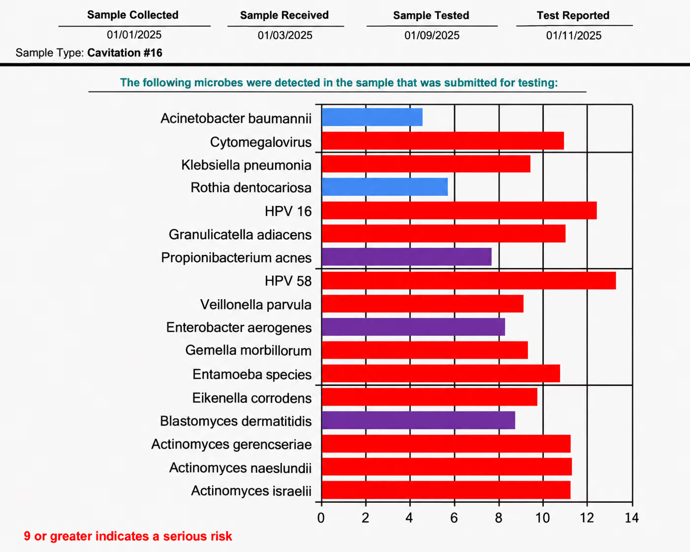
On a podcast, I mentioned that more than 90% of the old root canals I scan look concerning. That number comes from a specific group: patients seeking a second opinion because they already suspect a problem. It is not a statistic for every root canal.
The complex anatomy I mentioned earlier is one reason problems develop. Another study found that about 23% of treated premolars and molars had at least one missed canal. Those teeth were more than four times as likely to show inflammation around the root. This is one reason we refer to a specialist who works with a microscope.
How We Monitor a Treated Tooth
Because some problems remain silent, our usual protocol is to monitor existing root canals with a CBCT every 12 months. This is our practice philosophy rather than a universal guideline.
We use the smallest field of view and lowest dose that can answer the clinical question. If you have an older root canal that has not been evaluated in years, it is worth having it checked.
Worried About a Tooth?
If you’re experiencing discomfort or want to ensure your previous root canal is still healthy, contact our office to schedule an evaluation.
If the Tooth Needs to Come Out: Your Options
When extraction is the right choice, we talk through what should happen with the space afterward. The best option depends on your mouth, overall health, goals, and budget.
- Extraction, bone grafting, and an implant: This is my preferred path when the site is a good candidate. A bone graft can help preserve the area, and a qualified specialist places the implant because we do not place implants in our offices.
- Bridge: A fixed replacement supported by the neighboring teeth, which usually need to be reshaped. I recommend this less often due to having to cut down adjacent teeth.
- Removable partial: Sometimes called a flipper, this option has the lowest upfront cost but comes with tradeoffs in comfort and durability.
Costs vary by tooth and treatment. We provide procedure-specific pricing before you decide how to move forward.
What a Focused Exam With Us Looks Like
- When booking online, choose Emergency or Focused Dental Exam.
- This exam focuses on the specific tooth or concern rather than your entire mouth.
- Bring any existing X-rays and reports so we do not repeat imaging without a reason.
- If a CBCT scan or other X-ray is needed, we explain why and review the fee with you before taking the image.
If You Are in Pain Today
Urgent symptoms should never wait for a consultation. If you are hurting or suspect an abscess, call us promptly. We provide same-day care when we can do so safely.
An abscess needs treatment at its source. Medication may sometimes help control symptoms or a spreading infection, but it does not resolve the underlying dental problem on its own.
Go directly to the emergency room if you experience:
- Trouble breathing, speaking, or swallowing
- Swelling near the eye or spreading quickly across the face or neck
- Significant difficulty opening your mouth
- Fever accompanied by feeling severely ill
Get a Clearer Picture Before You Decide
If you have been told you need a root canal, or an older one is causing concern, we can help you understand what is happening. We’ll review your symptoms, examine the tooth, and look at your existing images before deciding whether a 3D scan would add useful information.